VIVO Pathophysiology
Other Topics
Goblet Cells
The talent of goblet cells is to secrete mucus, a viscous fluid composed primarily of highly glycosylated proteins called mucins suspended in a solution of electrolytes. Mucus serves many functions, including protection against shear stress and chemical damage, and, especially in the respiratory tree, trapping and elimination of particulate matter and microorganisms.
Distribution and Morphology
Goblet cells are found scattered among other cells in the epithelium of many organs, especially in the intestinal and respiratory tracts. In some areas, their numbers are rather small relative to other cell types, while in tissues such as the colon, they are much more abundant.
The image below is of a villus in the small intestine of a mouse. The section was stained using the periodic acid-Schiff technique, which stains glycoproteins, including mucins, bright purple.
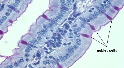
The large majority of cells covering the villus are absorptive epithelial cells, but several goblet cells are clearly visible. A similar type of situation is observed in bronchial and tracheal epithelium.
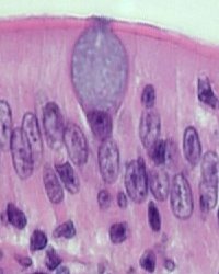
The name goblet cell derives from the characteristic shape of these cells in conventionally-fixed tissues: a narrow base and expanded apical portion that sometimes extends into the lumen. This morphology, as seen to the right in a section of cat small intestine (H&E stain), is known to be an artifact of fixation in which mucus-laden granules in the apical portion of the cell expand, causing the cell to balloon. If special precautions are taken during fixation, goblet cells are seen as cylindrical cells
Regardless of fixation, goblet cells have a distinctly polarized morphology. Their nucleus is at the base of the cell, along with organelles such as mitochondria, endoplasmic reticulum and Golgi. The remainder of the cell is filled with membrane-bound secretory granules filled with mucus.
Secretion of Mucus
Secretion of mucus from goblet cells is elicited primarily by irritating stimuli rather than in response to hormones. The lumen of the intestinal tract inevitably contains numerous irritants, and in the lung, such things as dust and smoke are potent inducers of goblet cell secretion.
Secretion of mucus is by exocytosis of secretory granules. Interestingly, goblet cells have two pathways for secretion:
- Constitutive or basal secretion: low level, unregulated and essentially continuous secretion. This pathway is dependent on cytoskeletal movement of secretory granules.
- Stimulated secretion: regulated exocytosis of granules in response to extracellular stimuli. This pathway provides an ability to dramatically increase mucus secretion
The mucus in goblet cell granules is condensed, but upon secretion, expands in volume tremendously and almost instantaneously (picture a pressurized can of shaving foam or whipped cream). In some systems studied, the mucin gel increases in volume 500-fold during a period of only 20 milliseconds!
A mechanism proposed for such rapid expansion of volume is as follows. Mucins are covered by abundant negative (polyanionic) charges, which, within secretory granules, are masked or "neutralized" by calcium ions. During exocytosis, a membrane pore opens to the outside of the cell, allowing calcium to diffuse out. This results in an extremely rapid phase change based on repulsion of polyanionic charges and hydration, leading to expansion of the mucin gel.
Pathophysiology
Goblet cells have the ability to differentiate into other cell types. A well-studied example of this is found in the respiratory tract, where goblet cells appear to be a progenitor to ciliated epithelial cells.
Increased numbers of goblet cells are observed in several disease states. Chronic brochitis and cystic fibrosis are examples of diseases in which goblet cell hyperplasia or metaplasia occurs.
References and Reviews
- Rogers DF: Airway goblet cells: responsive and adaptable front-line defenders. Europ Respiratory J 7:1690, 1997.
- Specian RD, Oliver MG: Functional biology of intestinal goblet cells. Am J Physiol 260:C183, 1991.
- Verdugo P: Goblet cells secretion and mucogenesis. Ann Rev Physiol 52:157, 1990.
Send comments to Richard.Bowen@colostate.edu