VIVO Pathophysiology
Other Topics
Ferritins and Hemosiderin
Iron presents a problem to living organisms - it is essential to nearly all cells but also is quite toxic. Iron is essential for its role in oxidation-reduction reactions and certain types of catalysis. Its toxicity comes from its propensity to form oxygen radicals that damage cells.
The solution that nature has evolved to meet the iron problem is a set of iron storage proteins that shield iron and prevent it from damaging other molecules, yet allow it to be released when needed.

Ferritins are class of iron storage proteins found in bacterial, plant and animal cells. They form hollow, spherical particles in which 2000 to 4500 iron atoms can be stored as iron(III) (i.e. Fe+++ ions). Depending on the organism, ferritin particles are roughtly 8-12 nm in diameter, with several channels that appear to mediate iron transport to and from the interior.
All ferritins are composed of 24 apoferritin monomers which associate to form a spherical particle. In mammalian cells, two types of ferritin monomers have been characterized (H and L types), which differ in the presence of certain residues that function to reduce ferric iron or facilitate mineralization of the particle with iron. Bacteria, including eubacteria and archaebacteria, have two types of ferritins: heme-containing bacterioferritins and heme-free ferritins. The structure of these molecules is very similar to those of animals and plants.
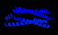
As shown in the figure above using a ribbon model, ferritin subunits are folded into four parallel helices with a fifth helix at roughly at 60 degree orientation.
In animals, ferritin is found not only inside cells, but circulating in plasma. Plasma levels of ferritin have sometimes been used as an index of iron storage deficiency.
The biosynthesis of ferritin is, as might be anticipated, controlled by the concentration of iron in the cell. Interestingly, such control is at the level of translation. A protein known as the iron regulatory protein (IRP) serves as an iron-binding molecule and sensor for cytoplasmic iron concentration. When iron concentration is low, IRP is not bound by iron and is capable of binding to the 5' end of the ferritin messenger RNA at a site called the iron regulatory element, a process which blocks translation of the mRNA. Conversely, when iron concentration is high, and the cell needs ferritin to protect itself, the IRP binds iron and becomes incompetent to bind the ferritin mRNA, leaving it available for translation.
Hemosiderin is another iron-storage complex. Its molecular nature remains poorly defined, but it is always found within cells (as opposed to circulating in blood) and appears to be a complex of ferritin, denatured ferritin and other material. The iron within deposits of hemosiderin is, at best, very poorly available to supply iron when needed. Hemosiderin is most commonly found in macrophages and is especially abundant in situations following hemorrhage, suggesting that its formation may be related to phagocytosis of red blood cells and hemoglobin.
References and Reviews
- Andrews SC: Iron storage in bacteria. Adv Microb Physiol 40:281-351, 1998.
- Chasteen ND, Harrison PM: Mineralization of ferritin: an efficient means of iron storage. J Struct Biol 126:182-194, 1999.
- Harrison PM, Arosio P: The ferritins: molecular properties, iron storage function and cellular regulation. Biochimica et Biophysica Acta 1275:161-203, 1996.
Send comments to Richard.Bowen@colostate.edu