
| Reproduction Index | Glossary |
|---|
|
Placentation in Ruminants (Cattle, sheep, ..) |
Ruminants comprise a large group of herbivores that include cattle, sheep, goats and deer. All of these animals, as described below, have cotyledonary placentae. Certain other animals, such as camels, while considered ruminants, but have diffuse placentae similar to what is seen in horses. ImplantationImplantation in ruminants is non-invasive and some authors prefer to use the term attachment. There is close attachment between embryonic membranes and the endometrium overlying caruncles at 5 weeks in cattle and 3 weeks in sheep. Shortly thereafter, the placenta is established. Gross Structure of the PlacentaRuminants have a cotyledonary placenta. Instead of having a single large area of contact between maternal and fetal vascular systems, these animals have numerous smaller placentae. The terminology used to describe ruminant placentation is:
Caruncles are oval or round thickenings in the uterine mucosa resulting from proliferation of subepithelial connective tissue. As shown in the image below, caruncles are readily visible in the non-pregnant uterus. Further, they are the only site in the uterus to form attachments with fetal membranes. Patches of chorioallantoic membrane become cotyledons by developing villi that extend into crypts in the caruncular epithelium. The image below shows caruncles in an incised non-pregnant sheep uterus (left) and cross sections through placentomes from a midgestation sheep pregnancy (right). Bovine placentomes looks similar, but have a convex appearance rather than the concave shape seen in sheep. 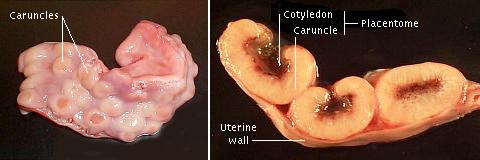 Pregnant sheep, goats and cattle have between 75 and 125 placentomes. Deer also have a cotyledonary placenta, but only 4 to 6 placentomes which are correspondingly larger. Further detail on gross structure of the ruminant placenta is presented below. | |
|
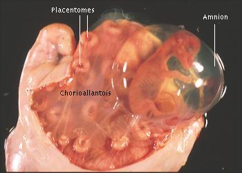
The image to the right shows an incised uterus from a pregnant sheep, roughly 50 days of gestation. The numerous button-shaped structures are placentomes, and the surfaces in view are actually cotyledons - the fetal side of the placentome. The slightly milky-looking membrane covering and between placentomes is the chorioallantois. The fetus is clearly visible inside the amnion. |
 |
| The image to the right shows a bovine conceptus dissected away from the uterus. The size of the chorioallantois relative to the amnion and fetus is evident. The cotyledons are readily observed; caruncles have been left behind with the uterus. Cattle almost always have a single fetus, and, although the fetus is located in one horn or the other, the large chorioallantosis fills both uterine horns, and placentomes are present throughout the uterus. | 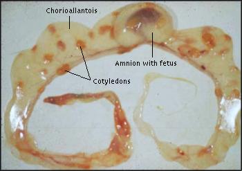 |
|
During parturition, there is substantial loosening of the cotyledonary villi from caruncles, and the placentomes expand laterally. After expulsion of the fetus and loss of fetal cirulation to the cotyledons, capillaries within the villi collapse, leading to a decrease in their size. The uterus contracts and the caruncular stock shrinks, further enhancing the separation of cotyledons from caruncles. In the normal case, fetal membranes with cotyledons are delivered within 12 hours of birth. There is no significant loss of maternal tissue, and therefore, ruminant placentation is considered non-deciduate. Microscopic Structure of the Placenta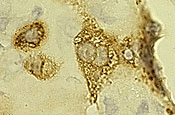
A prominent feature of the ruminant placenta is the presence of large numbers of binucleate cells. These cells arise early as part of the fetal trophoblast from cells that fail to undergo cytokinesis following nuclear division. They invade and fuse with caruncular epithelial cells to form small syncytia. Binucleate cells secrete the hormone placental lactogen. The image to the right shows a cluster of three binucleate cells immunostained (brown) for ovine placental lactogen (courtesy of R. Anthony). Ruminants basically have an epitheliochorial placenta, but because the uterine epithelium is modified by invasion and fusion of binucleate cells, its structure is generally referred to as synepitheliochorial. Prior to detailed study of these structures, it was thought that the maternal epithelium was eroded away, leaving trophoblast in contact with maternal connective tissue. The term syndesmochorial was used to describe this apparent structure, and is used in much of the older literature describing ruminant placentation. Placental TransportGeneral aspects aspects of placental transport are similar to that seen in other species. Immunoglobulins are not transported across the placenta from mother to fetus, and therefore, barring fetal infections, the young ruminant is born without circulating antibodies. Placental EndocrinologyThe major hormones of ruminant placentae are progesterone and other progestins, estrogens and placental lactogen. The sheep placenta produces enough progesterone that by roughly day 70 the corpora lutea can be removed and pregnancy will not be interrupted. In contrast, luteal progesterone is required throughout gestation in cattle and goats because their placentae secrete much smaller quantities of progesterone. In reality, a large amount of progesterone is synthesized by the goat placenta, but most is converted to a biologically inactive pregnane before secretion. The image below shows relative concentrations of progesterone and estrogens in pregnant sheep and cattle throughout gestation. The major species of estrogen is estrone.  The patterns of placental lactogen secretion is quite different in cattle and sheep. The bovine hormone is detected in maternal serum at about 4 months of gestation and remains low through parturation. In contrast, ovine placental lactogen is secreted in whopping quantities beginning at about day 50 and remains high through gestation. Placental lactogen also accumulates to high concentrations in the serum of fetal sheep, as shown in the image below.  | |
| Index of: Implantation and Development of the Placenta |
Last updated on August 8, 2000 |
| Author: R. Bowen |
| Send comments via form or email to rbowen@colostate.edu |