VIVO Pathophysiology
Functional Anatomy of the Endocrine Pancreas
The pancreas is an elongated organ nestled next to the first part of the small intestine. Its gross anatomy and the structure of pancreatic exocrine tissue and ducts are discussed in the context of the digestive system. The endocrine pancreas refers to those cells within the pancreas that synthesize and secrete hormones.
|
The endocrine portion of the pancreas takes the form of many small clusters of cells called islets of Langerhans or, more simply, islets. Humans have roughly one million islets. In standard histological sections of the pancreas, islets are seen as relatively pale-staining groups of cells embedded in a sea of darker-staining exocrine tissue. The image to the right shows three islets in the pancreas of a horse. | 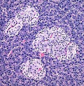 |
Pancreatic islets house three major cell types, each of which produces a different endocrine product:
- Alpha cells (A cells) secrete the hormone glucagon.
- Beta cells (B cells) produce insulin and are the most abundant of the islet cells.
- Delta cells (D cells) secrete the hormone somatostatin, which is also produced by a number of other endocrine cells in the body.
Interestingly, the different cell types within an islet are not randomly distributed - beta cells occupy the central portion of the islet and are surrounded by a "rind" of alpha and delta cells. Aside from the insulin, glucagon and somatostatin, a number of other "minor" hormones have been identified as products of pancreatic islets cells.
Islets are richly vascularized, allowing their secreted hormones ready access to the circulation. Although islets comprise only 1-2% of the mass of the pancreas, they receive about 10 to 15% of the pancreatic blood flow. Additionally, they are innervated by parasympathetic and sympathetic neurons, and nervous signals clearly modulate secretion of insulin and glucagon.
Send comments to Richard.Bowen@colostate.edu