VIVO Pathophysiology
Histology of the Adenohypophysis
The bulk of the adenohypophysis is pars distalis. That tissue is composed of winding cords of epithelial cells flanked by vascular sinusoids. In sections stained with dyes such as hematoxylin and eosin, three distinct cell types are seen among epithelial cells:
- Acidophils have cytoplasm that stains red or orange
- Basophils have cytoplasm that stains a bluish color
- Chromophobes have cytoplasm that stains very poorly
The figure below shows pars distalis from a cat at two magnifications (H&E stain).
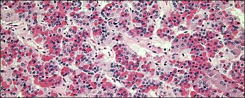
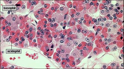
The differential staining pattern described above is a reflection of the type of hormonal content of the cells.
| Acidophils |
Cells that contain the polypeptide hormones:
|
|---|---|
| Basophils |
Cells that contain the glycoprotein hormones:
Due the high carbohydrate content of the hormones within acidophils, they also stain bright purple with PAS stains. |
| Chromophobes |
These are cells that have minimal or no hormonal content. Many of the chromophobes may be acidophils or basophils that have degranulated and thereby are depleted of hormone. Some chromophobes may also represent stem cells that have not yet differentiated into hormone-producing cells. |
Although classification of cells as acidophils or basophils is useful in some situations, specific identification of anterior pituitary cells requires immunostaining for the hormone in question.
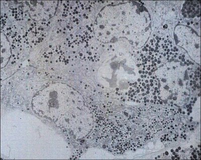
In addition to differential staining characteristics, the size of secretory granules varies among different types of cells in the anterior pituitary. Somatotropes and lactotropes tend to have the largest size granules. The electron microscopic image below of ovine adenohypophysis depicts cells with several densities and sizes of granules.
The portion of the adenohypophysis known as the pars tuberalis contains cords of epithelial cells and is filled with hypophyseal portal vessels. It reportedly contains gonadotropes and thyrotropes, plus other secretory cells of unknown function.
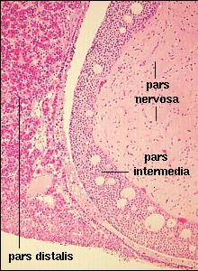
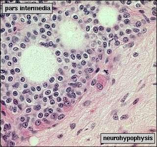
The pars intermedia is closely associated with pars nervosa and separated from the pars distalis by the hypophyseal cleft. This lobe of the pituitary shows considerable variation in size among species. It is small in man, but much larger in species such as amphibians. The pars intermedia contains large pale cells that often surround follicles filled with ill-defined "colloid". Melanocyte-stimulating hormone is the predominant hormone secreted by the pars intermedia. The images below show pars intermedia from a cat at low and higher magnification. The hypophyseal cleft is seen in the middle of the left image. In the right image, the three round, clear areas are follicles characteristic of this tissue.
Updated 2018. Send comments to Richard.Bowen@colostate.edu