VIVO Pathophysiology
Gross and Microscopic Anatomy of the Small Intestine
The small intestine is the longest section of the digestive tube and consists of three segments forming a passage from the pylorus to the large intestine:
- Duodenum: a short section that receives secretions from the pancreas and liver via the pancreatic and common bile ducts.
- Jejunum: considered to be roughly 40% of the small gut in man, but closer to 90% in animals.
- Ileum empties into the large intestine; considered to be about 60% of the intestine in man, but veterinary anatomists usually refer to it as being only the short terminal section of the small intestine.
In most animals, the length of the small intestine is roughly 3.5 times body length - your small intestine, or that of a large dog, is about 6 meters in length. Although precise boundaries between these three segments of bowel are not observed grossly or microscopically, there are histologic differences among duodenum, jejunum and ileum.
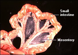
A bulk of the small intestine is suspended from the body wall by an extension of the peritoneum called the mesentery. As seen in the image to the right, blood vessels to and from the intestine lie between the two sheets of the mesentery. Lymphatic vessels are also present, but are not easy to discern grossly in normal specimens.
It is within the small intestine that the final stages of enzymatic digestion occur, liberating small molecules capable of being absorbed. The small intestine is also the sole site in the digestive tube for absorption of amino acids and monosaccharides. Most lipids are also absorbed in this organ. All of this absorption and much of the enzymatic digestion takes place on the surface of small intestinal epithelial cells, and to accomodate these processes, a huge mucosal surface area is required.
If the small intestine is viewed as a simple pipe, its lumenal surface area would be on the order of one half of a square meter. But in reality, the absorptive surface area of the small intestine is roughly 250 square meters - the size of a tennis court! How is this possible? At first glance, the structure of the small intestine is similar to other regions of the digestive tube, but the small intestine incorporates three features which account for its huge absorptive surface area:
- Mucosal folds: the inner surface of the small intestine is not flat, but thrown into circular folds, which not only increase surface area, but aid in mixing the ingesta by acting as baffles.
- Villi: the mucosa forms multitudes of projections which protrude into the lumen and are covered with epithelial cells.
- Microvilli: the lumenal plasma membrane of absorptive epithelial cells is studded with densely-packed microvilli.
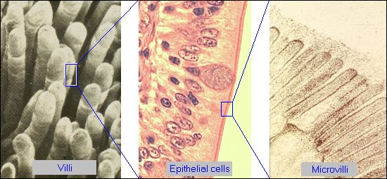
The panels below depict the bulk of this surface area expansion, showing villi, epithelial cells that cover the villi and the microvilli of the epithelial cells. Note in the middle panel, a light micrograph, that the microvilli are visible and look something like a brush. For this reason, the microvillus border of intestinal epithelial cells is referred to as the "brush border".
Most of the discussion on following pages focuses on enterocytes, the epithelial cells which mature into absorptive epithelial cells that cover the villi. These are the cells that take up and deliver into blood virtually all nutrients from the diet. However, several other important cell types populate the small intestinal epithelium:
- Enteroendocrine cells which, as part of the enteric endocrine system sense the lumenal environment and secrete hormones such as cholecystokinin and gastrin into blood.
- Goblet cells, which secrete a lubricating mucus into the intestinal lumen.
- Paneth cells, which provide an important anti-bacterial defense for the small intestine.
- Stem cells, which allow rapid and constant turnover of small intestinal epithelial cells, as described in the next topic.