VIVO Pathophysiology
Pancreatic Histology: Exocrine Tissue
The pancreas is surrounded by a very thin connective tissue capsule that invaginates into the gland to form septae, which serve as scaffolding for large blood vessels. Further, these septae divide the pancreas into distinctive lobules, as can clearly be seen in the image of mouse pancreas below (H&E).
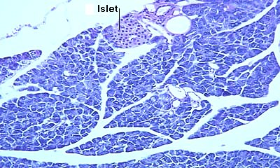
The large spaces between lobules seen in this image are a commonly-observed artifact of fixation.
The Acinus
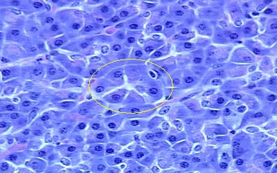
The exocrine pancreas is classified as a compound tubuloacinous gland. The cells that synthesize and secrete digestive enzymes are arranged in grape-like clusters called acini, very similar to what is seen in salivary glands. In standard histologic sections, most acini are cut obliquely, making it difficult to discern their characteristic shape. In the image of equine pancreas below, one fairly-good cross section through an acinus is circled; note the wedge-shapped cells arranged around a small lumen:
Pancreatic Ducts
Digestive enzymes from acinar cells ultimately are delivered into the duodenum. Secretions from acini flow out of the pancreas through a tree-like series of ducts. Duct cells secrete a watery, bicarbonate-rich fluid which flush the enzymes through the ducts and play a pivotal role in neutralizing acid within the small intestine. Pancreatic ducts are classified into four types which are discussed here beginning with the terminal branches which extend into acini.
- Intercalated ducts receive secretions from acini. They have flattened cuboidal epithelium that extends up into the lumen of the acinus to form what are called centroacinar cells.
- Intralobular ducts have a classical cuboidal epithelium and, as the name implies, are seen within lobules. They receive secretions from intercalated ducts.
- Interlobular ducts are found between lobules, within the connective tissue septae. They vary considerably in size. The smaller forms have a cuboidal epithelium, while a columnar epithelium lines the larger ducts. Intralobular ducts transmit secretions from intralobular ducts to the major pancreatic duct.
- The main pancreatic duct received secretion from interlobular ducts and penetrates through the wall of the duodenum. In some species, including man, the pancreatic duct joins the bile duct prior to entering the intestine.
Histologic features of the pancreatic duct system are illustrated in the following images:
A longitudinal section through an intercalated duct (Cynomologous monkey pancreas, H&E stain). The duct is running from upper left to lower right. Note the low cuboidal, almost squamous epithelium.
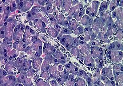
Section of equine pancreas (H&E stain) showing a longitudinal section through an intercalated duct emptying into an intralobular duct. Note the cuboidal epithelium in the intralobular duct.
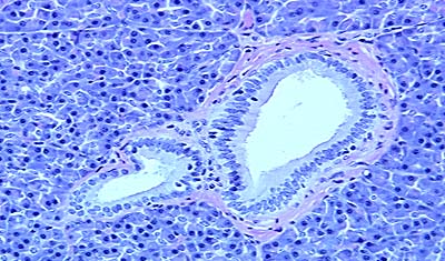
Small interlobular ducts (equine pancreas; H&E stain): note the columnar epithelium. A thin interlobular septum is seen running horizontally immediately above the duct.
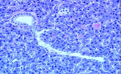
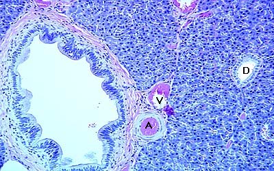
A low magnification image of equine pancreas (H&E stain) showing a large interlobular duct in association with a pancreatic artery (A) and vein (V). An intralobular duct (D) is seen on the right side.
Send comments to Richard.Bowen@colostate.edu