VIVO Pathophysiology
Hepatic Histology: The Lobule
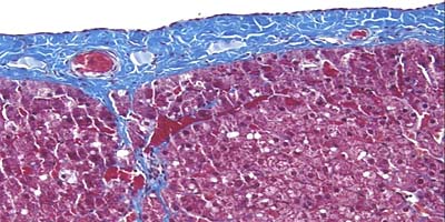
The liver is bounded by a connective tissue capsule which extends into its substance as highly branched septae. The afferent blood vessels and lymphatics follow this connective tissue highway throughout the liver. Efferent vessels traverse a route separate from connective tissue scaffolding.
In the section of equine liver below (Masson's trichrome stain), the capsule and septae are stained blue , while hepatocytes are magenta . Notice how the capsule extends as a septum into the liver about one-third of the way from left, immediately below a large capsular blood vessel.
The connective tissue septae invaginating from the capsule delineate hepatic lobules, the structural unit of the liver. Relative to other common species, the connective tissue surrounding lobules is particularly abundant and easy to identify in pig livers, as shown below in an H&E-stained section (Pass your mouse pointer over the image to confirm lobule boundaries).
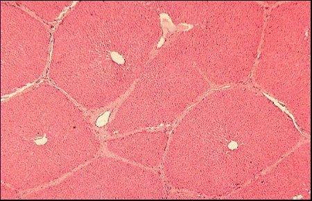
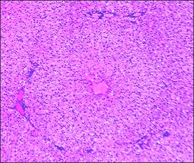
As you can observe above, a lobule is a roughly hexagonal arrangement of plates of hepatocytes radiating outward from a central vein (CV) in the center. Central veins are quite prominent and provide an easy means of orientation in sections of liver.
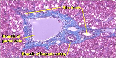
At the vertices of the lobule are regularly distributed portal triads (also known as portal tracts). Examination of a triad in cross section should reveal a bile duct and branches of the hepatic artery and hepatic portal vein. Due to plane of section, one can often observe more than one of each of these structures in a given portal tract or absence of one or more structures. Lymphatic vessels are also present, but are tough to see in standard paraffin sections, which is probably why it is not called a portal tetrad.
Lobules are almost impossible to miss in porcine liver, but one should also be able to recognize them in other species. Although the precise boundaries of lobules are sometimes difficult to discern, orienting on central veins and portal tracts allow "easy" identification. It goes without saying, of course, that the majority of lobules seen in a tissue section are not as "typical" as seen here and in other histology texts.
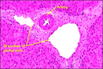
The following image is from a section of equine liver (H&E stain). See if you can picture the lobule, then move your mouse pointer into the image to confirm it.
Back to: Liver Histology: Introduction and Index ^
Send comments to Richard.Bowen@colostate.edu