VIVO Pathophysiology
Hepatic Histology: Extrahepatic Biliary System
Bile flows out of the liver through hepatic ducts, which join and extend as the common bile duct (also known simply as the bile duct) to traverse the wall of the duodenum and deliver bile into its lumen. In species with a gallbladder, the hepatic ducts join with the cystic duct, which conveys bile to and from the gall bladder.
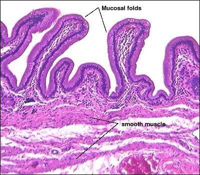
The gallbladder is a distensible sac and, when not distended, its mucosa is thrown into many folds. The lumen of the gallbladder is lined with a high columnar epithelium. The connective tissue wall contains abundant elastic fibers and layers of smooth muscle which predominantly run obliquely. The mucosa, connective tissue and muscularis of a canine gall bladder are seen below (H&E stain).
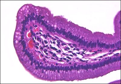
As seen below at a higher magnification, the epithelium of the gall bladder is quite uniform in appearance throughout the organ, and rests on a highly vascularized lamina propria. These epithelial cells are devoted to absorption of inorganic salts and water, and provide the mechanism for the gallbladder's ability to concentrate bile.
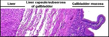
Between the smooth muscle layers and serosa is a thick subserosal layer of connective tissue. One face of the gallbladder is attached to the liver, and in that area, the connective tissue of the two organs is shared.
Back to: Liver Histology: Introduction and Index ^
Send comments to Richard.Bowen@colostate.edu