VIVO Pathophysiology
Architecture of the Liver and Biliary Tract
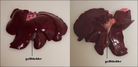
The liver lies in the abdominal cavity, in contact with diaphragm. Its mass is divided into several lobes, the number and size of which vary among species. In most mammals, a greenish sac - the gallbladder - is seen attached to the liver and careful examination will reveal the common bile duct, which delivers bile from the liver and gallbladder into the duodenum. The image below is of a liver from a dog, and illustrates these concepts. The panel on the left shows that aspect of the liver that faces the contents of the abdominal cavity. The right panel shows the flatter face of the liver which is in contact with the diaphragm.
Understanding function and dysfunction of the liver, more than most other organs, depends on understanding its structure. The major aspects of hepatic structure that require detailed attention include:
- The hepatic vascular system, which has several unique characteristics relative to other organs
- The biliary tree, which is a system of ducts that transports bile out of the liver into the small intestine
- The three dimensional arrangements of the liver cells, or hepatocytes and their association with the vascular and biliary systems.
The Hepatic Vascular System
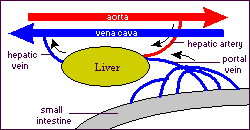
The circulatory system of the liver is unlike that seen in any other organ. Of great importance is the fact that a majority of the liver's blood supply is venous blood. The pattern of blood flow in the liver can be summarized as follows:
- Roughly 75% of the blood entering the liver is venous blood from the portal vein. Importantly, all of the venous blood returning from the small intestine, stomach, pancreas and spleen converges into the portal vein. One consequence of this is that the liver gets "first pickings" of everything absorbed in the small intestine, which, as we will see, is where virtually all nutrients are absorbed.
- The remaining 25% of the blood supply to the liver is arterial blood from the hepatic artery.
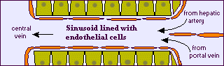
- Terminal branches of the hepatic portal vein and hepatic artery empty together and mix as they enter sinusoids in the liver. Sinusoids are distensible vascular channels lined with highly fenestrated or "holey" endothelial cells and bounded circumferentially by hepatocytes. As blood flows through the sinusoids, a considerable amount of plasma is filtered into the space between endothelium and hepatocytes (the "space of Disse"), providing a major fraction of the body's lymph.
- Blood flows through the sinusoids and empties into the central vein of each lobule.
- Central veins coalesce into hepatic veins, which leave the liver and empty into the vena cava.
The Biliary System
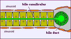
The biliary system is a series of channels and ducts that conveys bile - a secretory and excretory product of hepatocytes - from the liver into the lumen of the small intestine. Hepatocytes are arranged in "plates" with their apical surfaces facing and surrounding the sinusoids. The basal faces of adjoining hepatocytes are welded together by junctional complexes to form canaliculi, the first channel in the biliary system. A bile canaliculus is not a duct, but rather, the dilated intercellular space between adjacent hepatocytes.
Hepatocytes secrete bile into the canaliculi, and those secretions flow parallel to the sinusoids, but in the opposite direction that blood flows. At the ends of the canaliculi, bile flows into bile ducts, which are true ducts lined with epithelial cells. Bile ducts thus begin in very close proximity to the terminal branches of the portal vein and hepatic artery, and this group of structures is an easily recognized and important landmark seen in histologic sections of liver - the grouping of bile duct, hepatic arteriole and portal venule is called a portal triad.
Small bile ducts, or ductules, anastomose into larger and larger ducts, eventually forming the common bile duct, which dumps bile into the duodenum. A sphincter known as the sphinter of Oddi is present around the common bile duct as it enters the intestine.
The gall bladder is another important structure in the biliary system of many species. This is a sac-like structure adhering to the liver which has a duct (cystic duct) that leads directly into the common bile duct. During periods of time when bile is not flowing into the intestine, it is diverted into the gall bladder, where it is dehydrated and stored until needed.
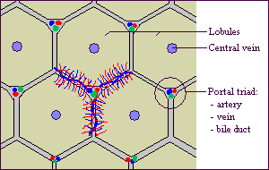
Architecture of Hepatic Tissue
The liver is covered with a connective tissue capsule that branches and extends throughout the substance of the liver as septae. This connective tissue tree provides a scaffolding of support and the highway which along which afferent blood vessels, lymphatic vessels and bile ducts traverse the liver. Additionally, the sheets of connective tissue divide the parenchyma of the liver into very small units called lobules.
The hepatic lobule is the structural unit of the liver. It consists of a roughly hexagonal arrangement of plates of hepatocytes radiating outward from a central vein in the center. At the vertices of the lobule are regularly distributed portal triads, containing a bile duct and a terminal branch of the hepatic artery and portal vein. Lobules are particularly easy to see in pig liver because in that species they are well deliniated by connective tissue septae that invaginate from the capsule.
Additional information on liver structure is presented in the sections on hepatic histology.
Send comments to Richard.Bowen@colostate.edu