VIVO Pathophysiology
Microanatomy of the Digestive Tube
Remarkably diverse and specialized processes take place in different sections of the digestive tract, but there is a fundamental consistency in the architecture of the tubular digestive tract. With few exceptions, the wall of the digestive tube from the mouth to the anus is composed of four basic layers or tunics.

Tunica serosa is the outermost covering of the digestive tube. In most of the digestive tract (stomach and intestines) it consists of a thin layer of loose connective tissue covered by mesothelium (a type of squamous epithelium that lines body cavities); within the peritoneal cavity, this structure is also referred to as visceral peritoneum.
In the abdominal cavity, the serosa on each side of the tube fuses together to form a suspensory structure called mesentery, which houses vascular and nervous supplies to the digestive tract and is continuous with the lining of the cavity. In regions outside of the abdominal cavity where the the digestive tube is essentially affixed to adjacent structures via its outer layer of connective tissue (esophagus and rectum), this tunic is referred to as tunica adventitia instead of tunica serosa.
Tunica muscularis endows the digestive tube with an ability to be motile. In most of the digestive tube, this tunic consists of two thick layers of smooth muscle. Muscle fibers in the inner layer are aligned circularly, whereas those in the outer layer have a longitudinal orientation.
This combination of circular and longitudinal smooth muscle gives the tube an ability to perform complex movements that squeeze and propel ingesta in the lumen. Between the inner circular and outer longitudinal layers of smooth muscle is another critical component of the digestive tract's nervous system - the myenteric plexus.
Tunica submucosa lies immediately beneath the mucosa, and is a layer of loose to dense connective tissue containing blood and lymphatic vessels. The submucosa also contains the submucous plexus, a critical component of the digestive tract's nervous system which provides nervous control to the mucosa.
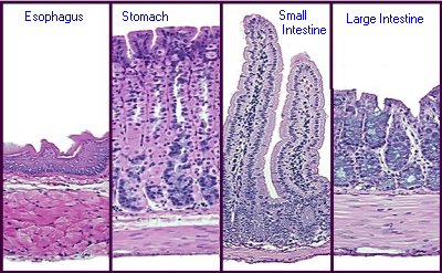
Tunica mucosa is the innermost layer of the digestive tube and lines the lumen. Among the four tunics, the mucosa is most variable in structure and function, endowing the tube with an ability to perform diverse and specialized digestive tasks along its length. Of critical importance in this regard are the epithelial cells that cover the mucosa and are thus in direct contact with the lumen. This epithelial cell sheet (lamina epithelialis) is distinctly different in different regions of the tract. Indeed, in most of the tract, several different cell types contribute to the epithelium, including cells dedicated to secretion, absorption or production of hormones.
These distinctive differences in architecture of the epithelium can be seen below in the micrographs of mouse digestive tube. The magnification of all four images is identical and the epithelial layer is oriented toward the top.
Beneath the epithelium, but still within the tunica mucosa is a layer - the lamina propria - of loose connective tissue through which course blood vessels and lymphatics that supply the epithelium. This layer also contains lymphatic nodules important to immune functions of the digestive tract. Finally, beneath the lamina propropria is a thin layer of smooth muscle (lamina muscularis mucosae) which permits the mucosa to dynamically move and fold.
Send comments to Richard.Bowen@colostate.edu